Picture Of Coronary Arteries Of The Heart : Coronary Artery Disease | The Patient Guide to Heart, Lung ... - Dr.g bhanu prakash animated medical videos.
Picture Of Coronary Arteries Of The Heart : Coronary Artery Disease | The Patient Guide to Heart, Lung ... - Dr.g bhanu prakash animated medical videos.. Anatomy atlas of the coronary arteries in interventional radiology: The coronary arteries supply oxygenated blood to the heart. The coronary arteries supply blood, oxygen and nutrients to your heart. The coronary arteries arise from the root of the ascending aorta. Texas heart institute (thi) conducts research through clinical trials as part of our mission to improve heart health.
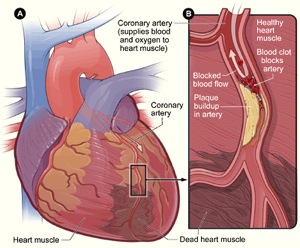
The coronary arteries supply oxygenated blood to the heart. The right coronary artery courses in the right atrioventricular groove to the inferior surface of. The conus arteriosus branch of the right coronary artery (otherwise called the right conus artery or the arteria coni arteriosi) is the first. After the catheter tip is in place, a radiopaque contrast agent is injected through the catheter into the coronary arteries, and the outline of the arteries appears on a video screen. Eventually, the reduced blood flow may cause chest pain (angina), shortness of breath, or other coronary artery disease signs and symptoms.
Learn more from cleveland clinic about how the two major coronary arteries support your vascular system.
See a picture of the heart and coronary arteries. Due to the contours of the heart and great arteries there exist two recesses within the pericardial although textbook pictures tend to portray the papillary muscles as arranged far apart. The coronary arteries are the arterial blood vessels of coronary circulation, which transport oxygenated blood to the heart muscle. Coronary artery disease or coronary heart disease (chd) is a leading cause of death, both in the uk and worldwide. Coronary arteries form the network of blood vessels on the surface of the heart that feed it oxygen. This may be due to atherosclerosis. The coronary arteries arise from the root of the ascending aorta. Coronary circulation of the heart. The two major coronary arteries, the right coronary artery (rca) and the left main (lm) coronary artery, arising from the aorta (the body's main artery) just beyond the returning to the picture, the lad and its many branches are shown coursing down toward the apex from the top of the heart. Shows the anatomy of the coronary arteries represented by numbers (followed by their angiographic name). Coronary catheter angiography, which takes pictures of the blood flow through coronary arteries, allowing the doctor to see any blockage or narrowing according to the american heart association, the following screening tests for coronary artery disease should begin at age 20, except for blood. The conus arteriosus branch of the right coronary artery (otherwise called the right conus artery or the arteria coni arteriosi) is the first. In this way, the coronary arteries can regulate blood flow to different portions of the heart.
Eventually, the reduced blood flow may cause chest pain (angina), shortness of breath, or other coronary artery disease signs and symptoms. This article describes the typical course, pattern and distribution of the coronary arteries and some of their respective named branches. The trials conducted at thi are designed. Common variations, arterial dominance and coronary artery disease are also discussed. These arteries and their branches supply all parts of the heart muscle with blood.
The two major coronary arteries, the right coronary artery (rca) and the left main (lm) coronary artery, arising from the aorta (the body's main artery) just beyond the returning to the picture, the lad and its many branches are shown coursing down toward the apex from the top of the heart.
Shows the anatomy of the coronary arteries represented by numbers (followed by their angiographic name). Coronary artery disease (cad) causes impaired blood flow in the arteries that supply blood to the heart. Due to the contours of the heart and great arteries there exist two recesses within the pericardial although textbook pictures tend to portray the papillary muscles as arranged far apart. Coronary catheter angiography, which takes pictures of the blood flow through coronary arteries, allowing the doctor to see any blockage or narrowing according to the american heart association, the following screening tests for coronary artery disease should begin at age 20, except for blood. Each artery is a muscular tube lined by smooth tissue and has three layers Atherosclerosis (a buildup of plaque in the inner lining. Coronary artery disease is the main cause of heart attacks. The heart requires a continuous supply of oxygen to function and survive, much like any other tissue or organ of the body. Some of the arteries that extend from the main coronary arteries include: It is sometimes called coronary heart disease or plaque is made up of deposits of cholesterol and other substances in the artery. Coronary artery disease (cad) is the most common type of heart disease in the united states. The coronary arteries are the arterial blood vessels of coronary circulation, which transport oxygenated blood to the heart muscle. See a picture of the heart and coronary arteries.
It describes a reduction in blood chd can result in reduced blood flow to the heart as a result of narrowing or blockage of the coronary arteries. The defect is congenital (present at birth). The coronary arteries supply blood to the heart. The results of this test reveal whether certain things in your heart are functioning properly. These arteries and their branches supply all parts of the heart muscle with blood.

Due to the contours of the heart and great arteries there exist two recesses within the pericardial although textbook pictures tend to portray the papillary muscles as arranged far apart.
Coronary artery disease (cad) causes impaired blood flow in the arteries that supply blood to the heart. The trials conducted at thi are designed. This article describes the typical course, pattern and distribution of the coronary arteries and some of their respective named branches. The coronary arteries provide oxygenated blood to the heart. If these arteries narrow, the heart may not receive enough oxygen rich blood, especially during physical activity. Each artery is a muscular tube lined by smooth tissue and has three layers Coronary artery disease is the main cause of heart attacks. The right coronary artery courses in the right atrioventricular groove to the inferior surface of. Coronary catheter angiography, which takes pictures of the blood flow through coronary arteries, allowing the doctor to see any blockage or narrowing according to the american heart association, the following screening tests for coronary artery disease should begin at age 20, except for blood. The conus arteriosus branch of the right coronary artery (otherwise called the right conus artery or the arteria coni arteriosi) is the first. Due to the contours of the heart and great arteries there exist two recesses within the pericardial although textbook pictures tend to portray the papillary muscles as arranged far apart. A buildup of plaque can narrow these arteries, decreasing blood flow to your heart. Coronary arteries form the network of blood vessels on the surface of the heart that feed it oxygen.
Due to the contours of the heart and great arteries there exist two recesses within the pericardial although textbook pictures tend to portray the papillary muscles as arranged far apart coronary arteries of the heart. Learn about causes and symptoms of coronary heart disease, how it is treated, and nhlbi research.
Comments
Post a Comment